1. Pemphigus Vulgaris
Disease generally begins in adolescent ages. Male/female ratio is equal. Initial lesions
are in the mouth almost always. Mouth blisters easily open and remain painful erosions.
  This localized phase lasts in 2-3 months. After this phase blister
formation is seen on the skin. Some blisters on the skin open and erosions
remain. One can see erosions dominantly on total body skin. But intact blisters
can be rarely seen on non-traumatized skin. Blisters typically appear on normal
skin without erythema and are fragile, slack and shrivel up. This localized phase lasts in 2-3 months. After this phase blister
formation is seen on the skin. Some blisters on the skin open and erosions
remain. One can see erosions dominantly on total body skin. But intact blisters
can be rarely seen on non-traumatized skin. Blisters typically appear on normal
skin without erythema and are fragile, slack and shrivel up.
 The
skin around the blister has normal appearance macroscopically, but intercellular
material has dissolved there and desmosomes are only binding structure.
When one press at the top of the blister, desmosomes disrupt by blister fluid
pressure. So, blister widens through the normal skin (Figure 1 at the left).
After hours of a frictional trauma on perilesional skin, a blister occurs on
the trauma site (Figure 2 at the left). One can easily pull the blister roof along the
normal skin, like a peach peeling (Figure 3 at the left). These three entities are
called as "Nikolsky phenomenon" and show acantholysis. The
skin around the blister has normal appearance macroscopically, but intercellular
material has dissolved there and desmosomes are only binding structure.
When one press at the top of the blister, desmosomes disrupt by blister fluid
pressure. So, blister widens through the normal skin (Figure 1 at the left).
After hours of a frictional trauma on perilesional skin, a blister occurs on
the trauma site (Figure 2 at the left). One can easily pull the blister roof along the
normal skin, like a peach peeling (Figure 3 at the left). These three entities are
called as "Nikolsky phenomenon" and show acantholysis.
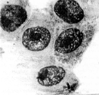 Smear material taken from the blister floor is mounted on a slide
and stained by Giemsa (Tzanck smear). One can see a typical keratinocytes with
large and dark stained nucleus and narrowed cytoplasm. These cells are also called
as "Tzanck cells" or "acantholytic cells". This
phenomenon shows acantholysis. Definitive diagnosis should be made by
intraepidermal blister formation and acantholysis. Smear material taken from the blister floor is mounted on a slide
and stained by Giemsa (Tzanck smear). One can see a typical keratinocytes with
large and dark stained nucleus and narrowed cytoplasm. These cells are also called
as "Tzanck cells" or "acantholytic cells". This
phenomenon shows acantholysis. Definitive diagnosis should be made by
intraepidermal blister formation and acantholysis.
Pemphigus vulgaris have the worst prognosis in pemphigus group.
Patients die in one year if they do not treat. Recent mortality rate is 25-40%
and mortality causes are the side effects of high dose of corticosteroids mainly.
|