PEMPHIGUSINTRODUCTIONPemphigus patients have IgG autoantibodies against intercellular material (cell membrane?). Antigen is a 130 kd polypeptide linked to plakoglobulin on binding sites. Patients have both of circulating antibodies (in serum) and deposited antibodies (in epidermis). Circulating antibodies can be shown by indirect immunofluorescence and deposited antibodies by direct immunofluorescence. Circulating antibody titration is correlated with disease activity. When disease improves titration decreases and when disease worsens titration increases.
Space, between keratinocytes is filled by fluid and blister formation occurs. Blister is situated in epidermis (intraepidermal blister). The roof of the blister consists of some superficial epidermal strata, so, roof of the blister is easily disrupted and blister opens. Secondary elementary lesion of the pemphigus blister is erosion. Erosion heals without scarring (because stratum basale is intact). There are four clinical types in pemphigus group:
|
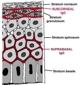
 IgG
immunoglobulins are deposited in Stratum spinosum, above stratum basale
(suprabasal) or in stratum granulosum (subcorneal). Deposited immunoglobulins are shown
in the figure at right. Intercellular material dissolves after antigen-antibody reaction.
Desmosomes try to bind keratinocytes weakly in this phase. But first trauma leads to
disrupt of desmosomes and keratinocytes becomes free. This entity is called as
acantholysis.